
Hematoxylin and eosin (H&E) are two special dyes that pathologists use to stain tissue samples, allowing them to be examined under a microscope. When tissue is removed from the body during a biopsy or surgery, it is processed and placed on glass slides for microscopic evaluation. However, without staining, the tissue appears nearly colorless, and the details of the cells and structures are hard to see.
Staining with hematoxylin and eosin adds color and contrast, making it easier for the pathologist to visualize the tissue organization and identify any signs of disease. Hematoxylin and eosin is the most common stain used in pathology and is often the first step in examining any tissue sample.
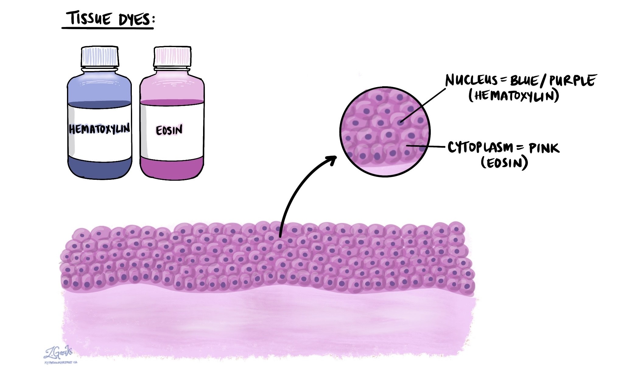
How does hematoxylin and eosin stain tissue?
Hematoxylin and eosin work together to stain different parts of the cells in two distinct colors, providing contrast between cellular structures:
-
Hematoxylin stains the nucleus of the cell a blue or purple color. The nucleus contains the cell’s DNA, which controls how the cell functions and divides.
-
Eosin stains the cytoplasm (the part of the cell outside the nucleus) and other supportive tissue structures a pink or red color. Eosin highlights proteins, connective tissue, and cell membranes.
By combining these stains, pathologists can see the size, shape, and arrangement of the cells and identify differences between normal and abnormal tissue.
How do pathologists use hematoxylin and eosin?
Pathologists use hematoxylin and eosin staining to examine tissue samples under a microscope and to identify signs of disease or injury. The two stains provide important contrast between different parts of the cell, making it easier to evaluate their structure and function.
-
Hematoxylin, which stains the nucleus blue or purple, is especially useful for evaluating changes in the size, shape, and color (chromatin pattern) of the nucleus. Since the nucleus contains the cell’s genetic material (DNA), changes in its appearance can give pathologists important clues about the health of the cell, including signs of cell damage, precancerous changes, cancer, or viral infections.
-
Eosin, which stains the cytoplasm and surrounding structures pink to red, helps pathologists assess the shape, amount, and texture of the cytoplasm, as well as changes in connective tissue, muscle, or blood vessels. Eosin is especially useful for identifying areas of cell death, tissue damage, or abnormal protein buildup.
Together, hematoxylin and eosin allow pathologists to see the overall organization of the tissue, detect abnormal cell growth, and identify patterns of disease. The hematoxylin and eosin stain is often the first and most important step in making a diagnosis and may be followed by other tests for more detailed information.
What other tests are used with hematoxylin and eosin?
While hematoxylin and eosin provide valuable information, some conditions require more detailed analysis. In such cases, pathologists may employ additional tests to aid in diagnosis or inform treatment.
Immunohistochemistry (IHC)
IHC uses antibodies that bind to specific proteins in the tissue. These antibodies are linked to dyes that make particular cells or proteins visible under the microscope.
IHC is commonly used to:
-
Identify the type of cancer (for example, whether it started in the lung or breast).
-
Detect the presence or absence of hormone receptors or tumour markers.
-
Determine whether cancer cells have spread to lymph nodes or other organs.
Special stains
These stains highlight specific features that are not visible with H&E alone.
Examples include:
-
Gram stain – used to identify bacteria.
-
PAS (Periodic acid–Schiff) – highlights carbohydrates, fungi, and mucus.
-
GMS (Grocott’s methenamine silver) – stains fungi black.
-
Ziehl-Neelsen stain – used to detect tuberculosis and other acid-fast bacteria.
Molecular tests
These tests look at the genetic material (DNA or RNA) in the tissue.
Molecular testing can:
-
Identify specific mutations that drive cancer.
-
Detect gene rearrangements or amplifications.
-
Help select targeted therapies for certain types of cancer.
Molecular testing is often used in cancers like lung cancer, thyroid cancer, and lymphoma, where treatment decisions depend on genetic findings.
Why is hematoxylin and eosin staining important?
H&E staining is the foundation of pathology. It allows pathologists to evaluate tissue in a way that is reliable, reproducible, and cost-effective. Without it, it would be nearly impossible to see important details in a tissue sample. Although newer and more advanced tests exist, they almost always build on the findings from the initial H&E-stained slide.
Questions to ask your doctor
-
What did the H&E stain show in my tissue sample?
-
Were any additional tests performed after the H&E stain?
-
Do I need further testing to confirm the diagnosis?
-
What do the results mean for my treatment?
We are proud to partner with:
![]()
Our work is generously supported by:
![]()